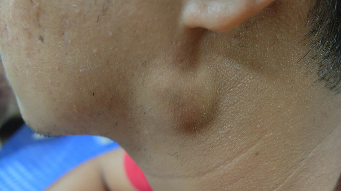
Even small or painless lumps in the jaw or cheek can signal a parotid problem. Noticing changes early allows timely assessment and treatment, improving outcomes and protecting facial function.
The parotid gland is one of the major salivary glands, located just in front of the ear and extending towards the cheek. It plays an important role in producing saliva, which helps with chewing, swallowing and overall oral health.
Parotid lumps are relatively common and can occur at any age. They develop when cells within the gland grow in an abnormal way, though the exact cause is not always clear. Most of these tumours are benign, accounting for about 80 percent of cases. However, a small number may be cancerous, which is why proper assessment is important.
Not all swellings are tumours. In some cases, salivary gland stones may block the ducts, leading to discomfort and recurrent infections. These conditions can feel similar at first, so a careful evaluation helps determine the cause and guide the next steps.
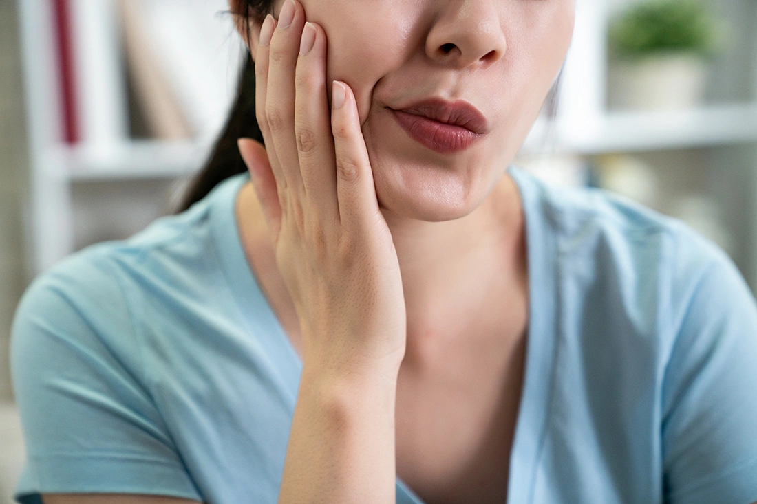
You may notice changes gradually or quite suddenly. Symptoms can vary depending on the underlying cause.
Common signs include:
Your consultation begins with a detailed discussion about your symptoms and medical history. This is followed by a clinical examination to better understand the nature of the swelling.
Imaging is often the next step. An ultrasound scan is commonly performed, and in many cases, a fine-needle sample is taken during the same session. This allows the cells to be examined under a microscope to determine whether the lump is benign or malignant. In selected situations, additional imaging such as CT or MRI scans may be recommended. Each investigation is discussed with you so you know what to expect and why it is needed.
Once a diagnosis is made, treatment can be planned with clarity. For most parotid tumours, surgical removal remains the main approach. This procedure, known as a parotidectomy, involves removing part or all of the gland depending on the condition.
Because the facial nerve runs through the parotid gland, surgery requires careful planning and precise technique. The goal is to remove the tumour completely while preserving facial movement as much as possible. In some cancer cases, additional treatment such as radiotherapy may be advised after surgery to reduce the risk of recurrence.
Not all procedures are the same. The recommended approach depends on the size, location and nature of the tumour.
This involves removing the outer portion of the gland where most tumours develop. It allows the tumour to be removed while preserving deeper structures, including the facial nerve.
When a tumour involves deeper areas or is more extensive, the entire gland may need to be removed. This approach is carefully planned to manage the condition effectively.
Sometimes referred to as an extracapsular dissection, this procedure removes only the affected portion of the gland, sparing as much healthy tissue as possible. It is typically reserved for small, benign and mobile lumps located in the tail of the parotid gland.
If a tumour recurs after a previous surgery, a revision parotidectomy may be necessary. This is more challenging than a primary surgery due to the presence of scar tissue, which can make the facial nerve harder to identify.
Beyond standard parotidectomies, other specific interventions may be required depending on the diagnosis:
If a lump is confirmed to be malignant, the surgery may need to be more extensive, potentially involving the removal of lymph nodes in the neck (neck dissection) to prevent the spread of cancer cells.
Fluid-filled sacs (cysts) can sometimes form in the gland, causing swelling and risk of infection. Surgery effectively removes the cyst wall to prevent recurrence.
Stones can block saliva flow and lead to pain or infection. If less invasive methods are not suitable, surgery may be considered.
Parotid surgery is generally safe when performed by an experienced surgeon. However, as with any procedure, there are some risks, which will be discussed with you beforehand.
Possible complications include:
Recovery after parotidectomy is generally straightforward for most patients. You can typically expect a hospital stay of one to three days to ensure you are healing well. Sutures are typically removed after about a week. As healing progresses, you can gradually return to your usual routine, with most daily activities resuming within two to three weeks.
Each recovery journey is different, but the aim is always to help you heal well while maintaining comfort and function.
A superficial parotidectomy involves removing only the outer lobe of the gland that sits above the facial nerve. In contrast, a total parotidectomy requires the removal of both the superficial and deep lobes. The choice of procedure depends entirely on where the tumour is located relative to the facial nerve.
Not every lump in the parotid gland is cancerous, and it can be difficult to tell by touch alone. A clear diagnosis involves a combination of clinical examination and advanced imaging, such as MRI or CT scans. A fine-needle aspiration biopsy is then used to analyse the cells. This thorough approach helps the specialist determine the nature of the lump and plan the most appropriate treatment.
The cost of parotid gland surgery can vary depending on the procedure (superficial or total parotidectomy), whether the tumour is benign or malignant, and whether additional reconstruction or nerve repair is needed. During a consultation, patients receive a personalised cost estimate and guidance on insurance coverage to ensure transparency and clarity.
Need clarity and guidance for cancer care?
Please reach out to us.
